CT Scan Guided Planning for Dental Implant Surgery – Houston, TX
High-Tech Surgical Plans
for Maximum Efficiency
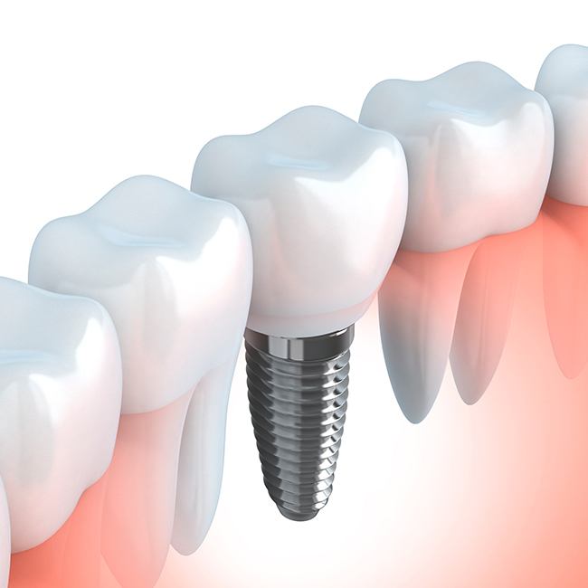
At Piney Point Oral & Maxillofacial Surgery, our oral surgeons use several current technologies that allow proper planning and dental implant placement for teeth that can augment surgical results tremendously. CT scans and computer planning help the surgeon place the dental implants with a high degree of precision and accuracy. This technology translates into a better outcome, improved cosmetic results, and potentially lifetime longevity of the implants.
What is CT Scan Guided Planning?

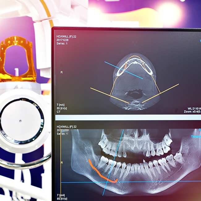
With our cone beam CT scan along with 3-dimensional treatment planning, dental implants can be placed in the exact position required to support the overlying crown or other prosthesis. Dental implant planning through the use of the CT scan allows the surgeon to look in the area of interest ahead of time to evaluate if the area needs to have additional bone grafting or if there is a problem with surrounding anatomy. The CT Scan does so by providing exact dimensions of the bone and surrounding structures such as nerves and sinuses, so dental implant placement can be done with accuracy and safely.
Three Stages of CT Guided Dental Implant Planning

When using computed tomography to plan an implant treatment, there are a number of steps we need to complete. Thanks to the state-of-the-art technology we employ, we can complete each of these steps relatively quickly and with high accuracy in order to ensure a successful surgery down the line.
Diagnostic CT Scan

Diagnostic CT scans can be completed right in the comfort of our oral surgery office. It is fast, safe, and very comfortable. First, a special splint with radiographic markers is made before taking the CT scan. This splint is placed in the patient’s mouth while the CT scan is being obtained to demonstrate the required position of the dental implant and aid the oral surgeon in treatment planning. The CT scan images with the markers already captured, will demonstrate the precise position of the dental implants, the exact size of the bone, as well as location and proximity of vital structures such as nerves and sinuses. A CT scan may also be done without the radiographic marker. In this case, it can be used to measure existing bone and evaluate other structures in proximity. The radiographic marker (also known as CT prosthesis) provides additional references for the surgeon and the restorative dentist when planning treatment.
Three-Dimensional Treatment Planning

Next, the CT data is reconstructed into a 3-dimensional computer model. With special software, the surgeon can now position the dental implants in the desired locations - all virtually. The correct width and length of the implants are selected to assure safe placement with respect to surrounding nerves and sinuses. This 3-dimensional model can be easily shared with the restorative dentist to make sure they are positioned with precision for the support of future prosthesis.
Surgical Guide

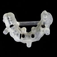
The computer dental implant work-up is then used to fabricate plastic surgical guides which will allow the surgeon to place the dental implants, according to the 3-dimensional virtual work-up. The surgical guides are made to fit the patient’s mouth with precision, allowing the surgeon to position the implants exactly as planned.
When is CT Guided Planning Recommended?

CT-guided dental implant planning is recommended in patients with full mouth implant restorations, those with high esthetic challenges, or patients with significant bone loss. CT-guided planning is not recommended in all patients because conventional planning can also lead to a successful result in many cases. But when a patient presents with some complexity, CT scans prove to invaluable to allow accurate imaging of the boney anatomy, which leads to accurate placement of the dental implants.

